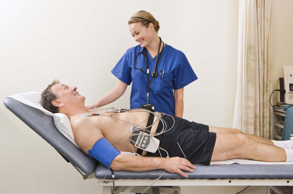
An electrocardiogram, or ECG, is a non-invasive test used to diagnose heart conditions. Performed in hospitals and physician’s offices, it’s among a medical assistant’s most exciting responsibilities. It’s a simple yet technical task that requires both people skills and sound clinical judgment. In a cardiology practice, a medical assistant interested in heart health with an aptitude for science can become an ECG technician.
What is an ECG?
Developed by Dutch doctor Willem Einthoven in 1903, the ECG maps electrical activity in the heart. As voltage flows through each of its four chambers, two upper and atria and two lower ventricles, the electrocardiograph captures its movement and records it as a wave pattern called a tracing. By measuring changes in the waves, doctors can tell how the heart is functioning.
How Does an ECG Work?
An ECG gathers and amplifies data from 12 leads applied to the body surface. Each lead records electrical impulses from a different angle, giving the physician a three-dimensional view of the heart. Tracings may be recorded on paper or stored digitally.
A standard ECG is performed with the patient at rest, but to evaluate symptoms such as chest pain that occur only during activity, the doctor might order a stress test, an ECG done while exercising, or a Holter monitor. Holter monitors are palm-size ECG devices that patients wear at home for periods of 24 hours or more to catch occasional irregularities in heart rate or rhythm.
Components of an ECG
ECGs record heartbeats as a wave. Each wave has five distinct features corresponding to different phases of heart function. Peaks and valleys in the waveform are labeled with letters; P, Q, R, S and T; to provide reference points for measurements. Groups of letters are also assigned to describe complexes and segments, the measurements between individual wave features.
P Wave
The P wave begins when both atria are filled with blood, and the SA node, a bundle of impulse-generating fibers in the wall of the right atrium, fires, causing them to contract. The SA, or sinoatrial node, is the heart’s natural pacemaker. Abnormalities suggest enlargement of the atria or failure of the node.
Q Wave
Q waves measure electricity as it travels through the cardiac septum. Irregularities suggest heart damage.
R Wave
On a normal ECG, the R wave is the tallest part of the waveform. It reflects the contraction of the left ventricle, the chamber that pumps blood into general circulation. Changes in R waves are evaluated as part of the QRS complex.
S Wave
The S wave is generated when the lower part of the heart muscle depolarizes, and the right ventricle contracts, sending blood to the lungs. Abnormal S waves may signal a pulmonary embolism.
T Wave
T waves mark relaxation of the ventricle and the end of the cardiac cycle. Abnormalities may not be pathological, but in a greater context, could reflect mitral valve damage, electrolyte abnormalities and heart enlargement.
PQ Segment
The PQ segment is the interval between the P and Q Waves. Shorter than average durations suggest Wolff-Parkinson-White Syndrome, the presence of a rare extra electrical pathway in the heart. Longer than average segments are the result of signal conduction impairments.
QRS Complex
The combination of the Q, R and S waves is called the QRS complex. Irregularities suggest defects in the heart’s electrical conduction system.
ST Segment
The amplitude of the ST segment reflects the time it takes for the ventricles to contract fully. It’s used as a baseline against which to measure other parts of the waveform. It’s among the most critical measures. Depressions in the ST segment reflect ischemia, a drop in the heart muscle’s blood supply caused by an obstruction. ST-elevation signals a heart attack.
Doctors consider the frequency, shape and size of each wave feature, segment and complex when evaluating ECGs.
What Do Doctors Learn from ECGs?
An electrocardiogram is only one diagnostic test used to evaluate heart function, but it can detect a range of serious cardiac disorders, including:
Dysrhythmias — irregularities in heart rate and rhythm
Ischemia — decreased blood supply to the heart muscle causing the chest pain known as angina
Cardiomyopathy — thickening of chamber walls due to heart failure or untreated hypertension
Electrolyte imbalances — high or low levels of ions that regulate most bodily functions
A history of heart attacks — the death of heart muscle caused by coronary artery blockages, such as blood clots or atherosclerotic plaques
Doctors order ECGs to rule out cardiac conditions as the cause of symptoms, including:
- Shortness of breath
- Lightheadedness
- Fatigue
- Chest Pain
Early diagnosis of heart disorders increases the chances they can be treated successfully.
Performing ECGs
Medical assistants are trained to perform ECGs from start to finish with only distance supervision. Steps include equipment preparation, patient screening, education and consent.
Equipment Preparation
Before doing an ECG, the medical assistant or technician completes quality control checks on equipment to ensure it functions correctly. Electrocardiographs are electrical devices, so other appliances that produce current could interfere with its function. Air conditioners, fans and cell phones should be turned off.
Patient Screening
ECGs are non-invasive, but for the best results, patients should lie flat on the exam table. However, because many people with heart disease have difficulty breathing in the supine position, it’s not possible for everyone. By pre-screening patients for physical limitations that could impact testing, medical assistants can make accommodations in advance and smooth the testing process.
Education and Consent
Patients have a right to know what to expect from an ECG, so medical assistants explain how the test is performed and answer questions before beginning. Precautions such as removing metal jewelry or body piercings that could interfere with readings are reviewed while patients change into a hospital gown or other loose-fitting clothing. Singed consent for testing may be required.
Performing the ECG
The actual ECG process is simple. The medical assistant places electrode pads on the patient’s body, avoiding placement over bones or broken skin, and attaches the color-coded leads. It might be necessary to shave hair and cleanse the skin for the pad adhesive to stick.
The electrocardiograph is turned on and programmed with the patient’s information. The medical assistant then asks the patient to take a deep breath and hold it while remaining still until the test is complete, it only takes a few seconds.
While medical assistants don’t interpret ECGs, they quickly learn which abnormalities could indicate an impending health crisis. Unexpected results should be reported to a doctor for evaluation before removing the electrodes.
ECG Aftercare
Once the test is complete, the medical assistant removes the electrodes and pads. Patients should be told when they can expect results and who they can call with questions.
The exam room is then sanitized for the next patient. While some general practitioners occasionally do screening ECGs in the office, a busy cardiology practice could do dozens per day, efficiency matters for a medical assistant working as an ECG technician.
Skills for An ECG Technician
Performing ECGs requires both practical and soft skills. The most important skills are compassion, attention to detail, communication, physical stamina, critical thinking, time management, and a commitment to learning.
Skill #1: Compassion
Medical assistants work with physically and emotionally sensitive people from all age groups and cultural backgrounds. While ECGs are typically well-tolerated, they can still cause anxiety for children, the elderly or patients from cultures unaccustomed to medical technology.
Since patient satisfaction is the focus of everything healthcare providers do, a gentle approach rooted in a deep sense of compassion is necessary for success.
Skill #2: Attention to Detail
Taking an ECG is straightforward, but each small step is precise, attention to detail is critical. A single misplaced lead can significantly alter results while missing subtle abnormalities can delay vital follow-up care.
Skill #3: Communication Skills
The ECG process requires giving and receiving large volumes of information, mixed messages can impact the process. Medical assistants should feel comfortable conversing with both patients and physicians. Active listening is a plus.
Skill #4: Active Listening
Active listening is a therapeutic technique in which an ECG technician focuses thoughtfully on what patients say both verbally and non-verbally. Reflecting statements clarifies the speaker’s intent, and interpreting body language gives communication more depth and accuracy.
Skill #5: Physical Stamina
ECG technicians spend most of the day on their feet. There is a need to bend, twist and stoop to place electrodes and assist patients with mobility challenges. Energy and physical stamina are essential.
Skill #6: Critical Thinking
Critical thinking is the ability to analyze facts and come to reasonable conclusions. For an ECG technician, it’s a necessary part of making clinical judgments. Medical assistants learn all they need to know in school to handle patient’s needs, but experience and critical thinking are what help them to prioritize tasks, manage changing circumstances and identify risky clinical situations.
Skill #7: Time Management Skills
Performing ECGs is an exacting process requiring a sense of calm and focus, but medical offices are fast paced. The ability to multitask and manage time effectively by using each minute to its fullest potential helps keep the schedule on track and minimizes stress.
Skill #8: A Commitment to Learning
Healthcare is always evolving. While vocational school graduates have all the skills they need to perform electrocardiograms, becoming certified as an ECG technician is another way job applicants can demonstrate their skills. Certification isn’t necessary to enjoy a full career, but it reflects a commitment to the medical field that employer’s value.
Final Thoughts
Vocational school programs prepare students to handle a broad array of clinical and administrative responsibilities in a medical setting, it’s a rewarding role. But for graduates with a love of science and technology, the option to work as an ECG technician is one worth considering.
Did learning how to become an ECG technician interest you? Ready to learn more about becoming a medical assistant? Meridian College offers hands–on Medical Assistant training from experienced school faculty who know how to prepare you for the daily challenges you’ll face on the job. From assisting doctors with patients to important administrative tasks, our experienced Medical Assistant program teachers will train you for a rewarding new career.
In addition to receiving training from school instructors with real-world experience, you will also complete a school externship in a physician’s office, clinic or related healthcare facility under the supervision of a physician, nurse or health services professional to further develop your skills.
Contact Meridian College today to learn more about becoming a medical assistant.